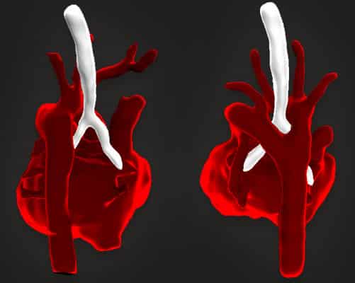
Scientists from Evelina London and King’s College London have developed a new method using MRI to produce detailed 3D images of the fetal heart. This will improve the diagnosis of congenital heart disease in babies before they are born.
Tiny, fast moving, unborn babies can be difficult to diagnose with ultrasounds, meaning it can be hard to see abnomalites in the blood vessels around their hearts.
In a paper, published in the Lancet, the team from Evelina London and King’s College London write about how they successfully used a new computer processing method for when cogenital heart disease is suspected. Their research allowed standard MRI images, which are often unclear because of the moving baby, to become clear three-dimensional models. The 3D models we used for 85 pregnant women and provided reliable, high-resolution information.
John Simpson, professor of paediatric and fetal cardiology at Evelina London, said: “Three dimensional MRI revolutionise the type of information we can obtain before babies are born. This impacts directly on care we provide after birth and provides new insights into structural heart defects before birth.”
Dr David Lloyd, clinical research fellow at King’s College London said: “Our hope is this approach will now become standard practice for the Evelina fetal cardiology team who make a prenatal diagnosis in 400 babies each year. This will also improve the care of over 150 babies each year who deliver at St Thomas Hospital with known congenital heart disease.”
Reza Razavi, professor of paediatric cardiovascular science and consultant paediatric cardiologist at Kings College London, said: “Application of this novel computing technology has enabled for the first time MRI scanning to really help with clarifying the diagnosis in a subgroup of babies, particularly when the vessels around the heart are involved.”
The team are now working to combine this 3D imaging with other advanced ultrasound and MRI techniques. They hope this may help them to understand why some babies go on to develop more severe forms of congenital heart disease than others.
This work is supported by the iFind project, which is exploring novel robotics, ultrasound, MRI and computing techniques to improve the prenatal diagnosis of fetal abnormalities.
Find out more about fetal cardiology at Evelina London.
News article and image source:
https://www.evelinalondon.nhs.uk/about-us/news-events/2019-news/20190325-images-before-birth.aspx